32
Vol. 66, No. 1 2015
Northeast Florida Medicine
Otolaryngology
The vHIT uses high speed infrared recordings of eye po-
sition to measure VOR driven eye movements in response
to short, rapid head accelerations. These head impulses
can be made in the planes of each SSC to estimate canal
specific VOR responses.
8,9
The vHIT can show two types of
abnormalities: weak VOR responses (decreased VOR gain)
or near normal eye movements accomplished by centrally
driven corrective saccades. The latter reflects brainstem/
cerebellar generated compensatory behaviors.
10,11
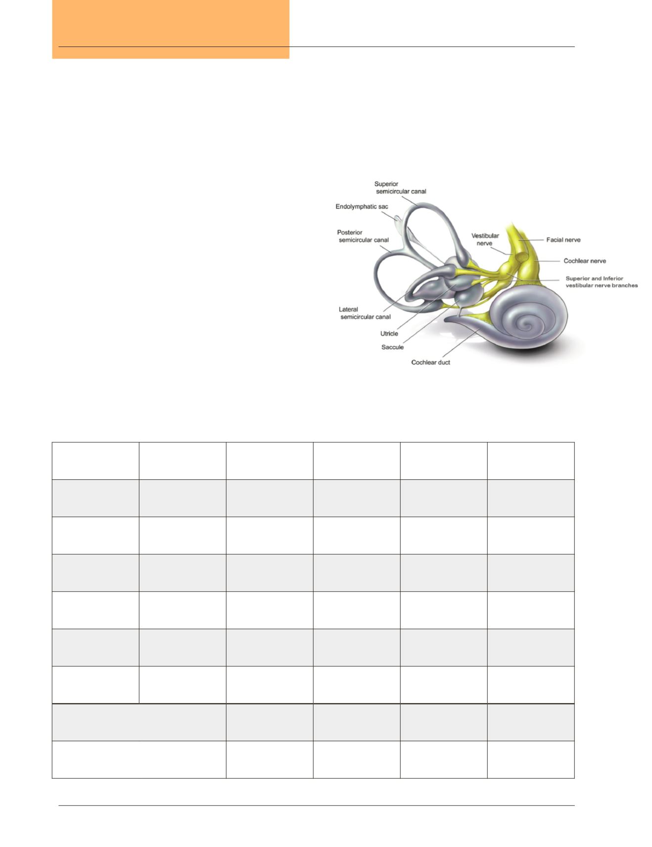
The value of vHIT and VEMP tests can be understood in
relationship to the innervation patterns themembranous lab-
yrinth. The three major branches of CNVIII innervate only
a few of the sensory structures in the membranous labyrinth
(Figure 2). By recognizing patterns of test results, specific
nerve syndromes can be appreciated.
11,12
These relationships
are shown in Table 2. When single nerve branches are in-
volved, recovery potential is high. When multiple branches
are involved, however, recovery will be sub-optimal, and the
risk of retro-labyrinthine disease increases.
1
A superior vestibular nerve syndrome is the most com-
monly encountered cause of acute vertigo in the clinic, after
Table 2.
vHIT, VEMP and audiogram test result patterns for various vestibular nerve syndromes.
Test
Structure Assessed Superior Nerve
Syndrome
Inferior Nerve
Syndrome
Superior/Inferior
Syndrome
Global Syndrome
(all branches
involved)
vHIT
Superior / Anterior
SSC
Abnormal
Normal
Abnormal
Abnormal
vHIT
Horizontal SSC
Abnormal
Normal
Abnormal
Abnormal
oVEMP
Utricle Otolith
Abnormal
Normal
Abnormal
Abnormal
vHIT
Posterior / Inferior
SSC
Normal
Abnormal
Abnormal
Abnormal
cVEMP
Saccule Otolith
Normal
Abnormal
Abnormal
Abnormal
Audiogram
Cochlea
Normal or Symmetric
Hearing Loss
Normal or Symmetric
Hearing Loss
Normal or Symmetric
Hearing Loss
Hearing Loss Greater
in the Involved Ear
Prognosis
Good
Good
Poor
Poor
Risk of CN VIII Tumor
Low
Low
Moderate
Very High
Figure 2.
Cranial Nerve VIII Innervation Pattern.
(Figure Copyright 2014, Mayo Clinic Foundation for
Medical Education and Research.)
Figures used with the permission of Mayo Foundation for
Medical Education and Research, all rights reserved


