Northeast Florida Medicine
Vol. 66, No. 1 2015
43
Otolaryngology
Another risk with any new and promising therapy is
over-utilization.We are all familiar with the adage, “when all
you have is a hammer, everything looks like a nail.” Because
of its advantages, balloon sinus procedures have been used
in acute sinusitis, headaches and other situations where its
efficacy may be unproven or even dubious.
4
These authors
have been performing balloon sinus surgery for years but
would still recommend a measure of restraint when recom-
mending this as an office or solo procedure.
Finally, balloon-assisted techniques are not effective
in treating nasal obstruction due to deviated septum or
enlarged turbinates. Since nasal obstruction is a common
presenting complaint in patients with chronic sinonasal
infection, an exclusively balloon-based technique (whether
in office or operating room) may not address all of the
patient’s complaints. Simply put, balloon-assisted sinus
surgery is not a panacea and is unlikely to eliminate the
need for additional, more established endoscopic sinus
techniques in the surgeon’s armamentarium.
Stereotactic Sinus Surgery
Another technique which has gained popularity and wide
acceptance is stereotactic or “image guided” sinus surgery.
Using special equipment in the operating room, thin-slice
axial CT scans are compiled into a three-dimensional model
which can be used to locate the position of instruments
within the head, much like a “GPS for the sinuses.”
5
Pros and Cons
There are several obvious advantages to image-guided
sinus surgery. Given the proximity of orbital, CNS and vas-
cular structures, image guidance is a helpful adjunct to the
high-definition endoscopy that is the mainstay of modern
sinus approaches. In addition to risk reduction, image guid-
ance may also help to elucidate difficult anatomy, such as in
cases of variant or small anatomy, revision surgery, polyposis
or severe mucosal disease. Image guidance also allows the
surgeon to target specific areas for surgical exposure or biopsy.
The disadvantages to the image guidance system (IGS)
primarily involve cost and availability. IGS equipment re-
quires the use of disposable cables and guides which add cost
to a patient’s surgical bill and may be superfluous in cases
of mild or limited sinusitis. Image guidance also requires
specially-formatted CT scans: if original sinus CTs are not
done with IGS in mind, scanning may need to be repeated
to create compatible images. Finally, due to its cost, IGS
may not be available in smaller hospitals or surgery centers.
Another potential disadvantage is the radiation exposure
from CT scans, currently the only imaging modality for
which stereotactic guidance is widely available. Concern over
correctly positioned. Another technique involves a balloon
which is placed through a small puncture above the gum
line into the maxillary sinus and then passed, inside-out
fashion, through the natural ostium into the nasal passage.
2
Pros and Cons
The potential advantages of balloon sinus surgery are
significant and attractive, in that the technique offers po-
tentially less scarring, bleeding, and postoperative pain than
more aggressive and traditional techniques. Balloon sinus
dilation can also be performed in an office or clinic setting,
often eliminating the need for general anesthesia.
3
As with many new surgical techniques there are potential
drawbacks, as well.The most obvious is that balloon-assisted
sinus surgery may not be appropriate or applicable in all
cases. Balloon-assisted surgery works most effectively when
there is a single bony ostium or outflow tract, such as with
the frontal or sphenoid sinus. The ethmoid sinuses, on the
other hand, are a group of small sinuses without a single
outflowchannel; to date, there has not been awidely-accepted
balloon solution to chronic ethmoid disease. Although data
exists to support its use in the maxillary sinus, there has been
mixed success at eliminating chronic maxillary disease with
balloon dilation alone.
4
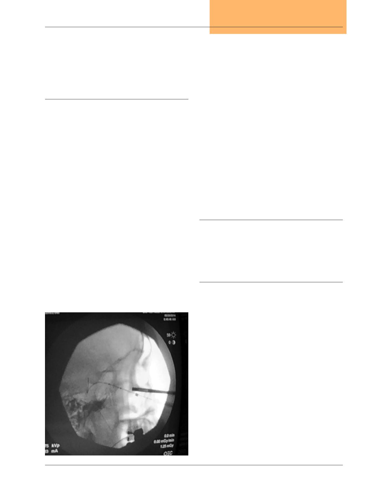
Figure 2.
Seldinger-type balloon dilation of sphenoid sinus (lateral
view) under fluoroscopy. Note soft-tip guidewire in
sphenoid sinus. Radiopaque marks along wire indicate
anterior and posterior margins of balloon to be inflated.


