Northeast Florida Medicine
Vol. 66, No. 1 2015
49
Otolaryngology
Often times, these symptoms, without an explainable
cause, will prompt imaging studies such as an MRI of the
brain, CT of the sinuses and temporal bones, carotid ul-
trasound, CT angiogram, and MR angiogram. Very often
these tests are normal or have incidental, unrelated findings
that do not reveal an underlying condition or diagnosis
attributable to the symptoms. Patients and providers are
often frustrated with a negative evaluation, leading to self-
doubt by the patient and concerns about the legitimacy of
the symptoms by the provider. Patients have usually been
referred to multiple specialists and have often been told, “I
cannot find anything wrong.”
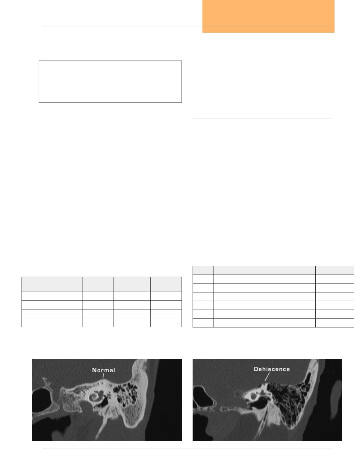
The key to diagnosis is the high resolution CT scan of the
temporal bones without contrast, with reconstructions in
the Poschl’s (Figures 2 & 3) and Stenver’s planes. (Table 1)
In Figures 4 & 5, the planes are perpendicular and parallel
to the superior semicircular canals. These reconstructions
are not typically done in the routine, high resolution CT
scan of the temporal bones. Standard high resolution CT
scans of the temporal bones without these specific planes of
reconstruction will overlook the dehiscence of the superior
semicircular canal.
Treatment
Current treatment involves either simple conservative
treatment or surgical intervention. There are not any medi-
cations that are known to be effective in relieving symptoms.
Most patients are content with observation and expectant
management once they know there is an abnormality that
explains their unusual symptoms. Once reassured of a specific
diagnosis and their symptoms are not occult manifestations
of a more sinister diagnosis, the symptoms are usually much
better tolerated.
The surgical management is evolving. There are two
primary surgical strategies: plugging the defect directly or
resurfacing the defect
7-11
.The surgical approaches for plugging
involve either a middle fossa craniotomy or a transmastoid
labyrinthotomy.The surgical approaches for resurfacing also
involve amiddle fossa craniotomy or a transmastoid approach.
- 64 Slice multi-detector scanner
- Collimation for acquiring data: 0.6 mm at 0.3 mm intervals
- Reconstruction in oblique sagittal and oblique coronal planes
(0.8 mm thickness at 0.2 mm intervals)
Table 1.
CT scan protocol Table 1. CT scan protocol
Table 2.
Comparison of surgical approaches
Table 3.
Outcome metrics
Surgical approach
Length
of stay
Superior canal
function
Operative
risks
Middle fossa plugging
2 – 5 days
Diminished Moderate
Middle fossa resurfacr
2 – 5 days
Preserved
Moderate
Transmastoid plugging
2 – 5 days
Diminished
Low
Transmastoid cartilage cap Outpatient
Preserved
Low
Name Description
Type
FGA Functional Gait Assessment (21)
Physical therapy
DHI
Dizziness Handicap Inventory (22)
ABC Activities-specific Balance Confidence scale (23)
GAD 7 Generalized Anxiety Disorder (24)
PHQ 9 Depression Questionnaire (25)
VAS Visual analog scale (Tullio, Hennebert, autophony)
Figure 4.
Normal superior semicircular canal, Stenver view.
Figure 5.
Dehiscent superior semicircular canal, Stenver view.
All Figures: Christine Gralapp ©


