16
Vol. 66, No. 3 2015
Northeast Florida Medicine
Endovascular Neurosurgery
The Development of
Cerebral Angiography
Endovascular technique requires that the operator see
what he is doing.The basis of almost all current endovascular
therapy is the cerebral angiogram. Moniz, a Portuguese
neurologist, is credited with the discovery of angiography.
4
Prior to this, the only method for imaging the brain was by
pneumoencephaolography, which introduced air directly
into the cerbrospinal space. This painful and cumbersome
method, developed by Dandy, had only limited utility for
imaging vessels.
4
Early attempts to inject contrast media
into arteries included an injection of the hand of a corpse
with a toxic amalgamof petroleum, quicklime andmercuric
sulfide, and several reports of injection of lipiodol into
limbs had failed to provide images.
4
Moniz believed that
if he could introduce a radiopaque material that would
concentrate in the brain, the brain could become visible on
x-rays.
4
In his first attempts, Moniz gave patients large oral
doses of bromides. However, the brain was not opacified
on subsequent radiographs. He then performed direct
injections of bromide solution into the carotid arteries of
dogs and into the vessels of cadaver heads that he arranged
to have delivered to his laboratory in taxicabs.
4
He eventually
obtained very crude images of the cerebral vessels.
The first reported successful cerebral arteriogram took
place in 1927. Moniz performed surgical exposure and
temporary ligation of the carotid artery and injected a
solution of 25 percent sodium iodide.
4,5
Interestingly, while
hailed as a great advance by the neurology community at
the time, the technique was somewhat slow to be adopted.
Still, by the 1950s, cerebral angiography had become the
dominant imaging modality for the brain, and it remained
so until the advent of computed tomography (CT) scanning
in the 1970s.
6
For many years, cerebral angiography
was the only reasonable imaging method to look at the
human brain. It was performed mostly by neurosurgeons,
and generally by surgical cutdown or later direct carotid
cannulation. Gazi Yasargil, a neurosurgeon recognized as
one of the fathers of the microsurgical treatment of cerebral
aneurysms, is reported to have performed over 10,000
angiograms between 1953 and 1964.
7
Moniz received the Nobel Prize in 1949 – but not for his
workwith angiography. He was honored for the development
of the frontal lobotomy procedure.
8
At the time, with no
medicines to control severely mentally ill patients, this
procedure was widely adopted to control unruly psychiatric
patients. Angiography has clearly been a far more significant
and lasting contribution to modern medicine.
Cross-sectional imaging techniques such as CT and
magnetic resonance imaging (MRI) have supplanted
cerebral angiography as a diagnostic tool for most brain
imaging. However, angiography remains the gold standard
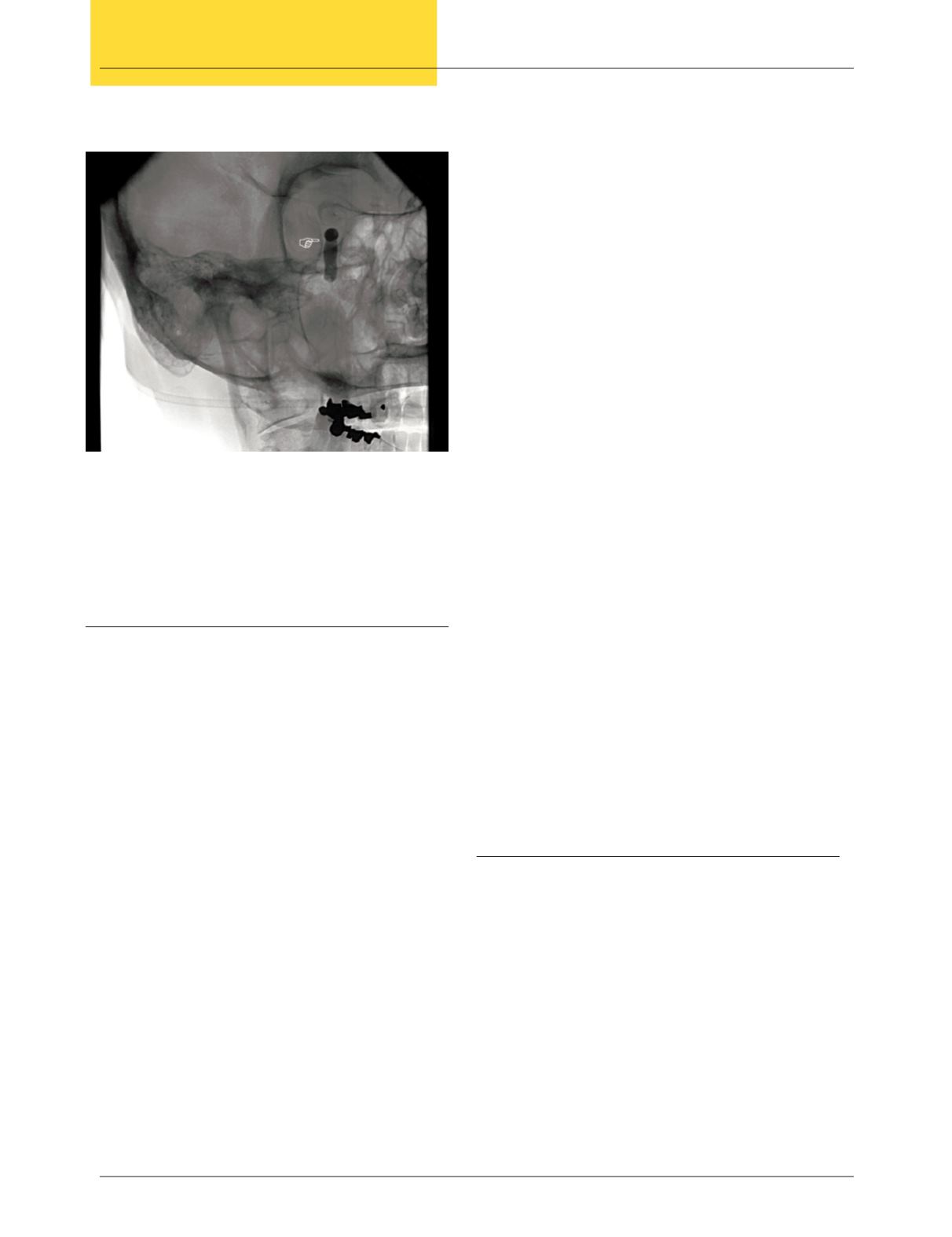
for vascular imaging. In addition, advances in angiography,
including digital subtraction techniques, “road map” real-
time image overlays, and rotational arteriograms, allowing
three-dimensional reconstruction, have become invaluable
in both planning interventional procedures and in providing
precise intraprocedural images to guide treatment. (Figure 1)
The Birth of
Modern Endovascular Technique
Reports of efforts to embolize cerebral vascular lesions
appeared soon after Moniz’s description of the cerebral
angiography procedure. However, these procedures hardly
resemble what we would consider true “endovascular”
procedures today. Most were performed in the context of
a surgical procedure, or involved surgical exposure of the
cervical carotid artery to place a catheter. Embolic agents
were often introduced directly into the vascular lesion,
or were released into the cervical carotid artery, far from
the target lesion. The technology did not exist to advance
catheters into the distal cerebral vasculature. In 1930, Brooks
described embolization of a carotid cavernous fistula (CCF)
by placing thin pieces of muscle in the internal carotid artery.
The pieces were then carried to the fistula. However, more
Figure 1: Anterior communicating artery aneurysm
A. Aneurysm sac with measurements
B. Anterior Cerebral Artery (parent vessel for aneurysm)
C. Ventricular drain (for subarachnoid hemorrhage)


