Northeast Florida Medicine
Vol. 66, No. 3 2015
17
Endovascular Neurosurgery
recent reviews of the article suggest that the muscle may
have been placed by a direct surgical procedure, as opposed
to delivered using arterial flow.
9
Still, the procedure framed
the concept, and it became the standard treatment for CCFs
until the development of detachable balloon catheters.
In 1941Werner et al. inserted silver wire into an aneurysm
during a transorbital craniotomy, and heated the wire to
80 degrees centigrade for one minute.
10
They reported
occlusion of the aneurysm sac. In 1965, Mullen reported a
series of 12 aneurysms treated with electrothrombosis using
stereotactic placement of the electrode needle through a
burr hole. It was clear that many still thought that there was
merit in less invasive strategies. In 1958, Luessenhop and
Spence described embolization of a large left hemispheric
arteriovenous malformation (AVM) with spheres of methyl
methacrylate ranging from 2.5 to 4.2 mm in diameter.
11
The
carotid was surgically exposed and the embolic material was
released into the left cervical internal carotid artery. The
internal carotid artery (ICA) was then ligated. Luckily, the
high flow of the AVM carried the embolic material to the
target. This proved that arterial flow could be used to deliver
embolic material in the brain. While the achievement was
groundbreaking, and actually remains the concept of how
we treat many AVMs today, it was clear that there had to
be a better way to deliver such material.
By 1950, cerebral angiography had become relatively
routine at largemedical centers, as a diagnostic tool. Accessing
the cervical arteries was not without risk. Generally, access
was through the carotid arteries, either by cut down or direct
puncture. The studies were performed through a large bore
needle puncture which left a large hole in the artery and
the risks of arterial wall damage were high. In 1951, Pierce
developed a polyethylene catheter for angiography.
12
This
could replace the much more traumatic and less flexible
angiography needles. Two years later, Seldinger developed a
technique for placing the flexible catheter into an artery over
a guidewire.
13
This reduced much of the risk involved with
percutaneous arterial punctures.His work also showed that all
arteries in the body could be reached from a femoral arterial
puncture. This eliminated the more risky carotid artery
punctures. Angiography, as a technique for both diagnosis
and therapy, suddenly became much safer to perform, and
could be much more widely applied.
12,13
The Rise of Endovascular Devices
The era of catheter-based endovascular therapy for the
intracranial compartment really started to take off in the
late 1960s and early 1970s. Advances in materials, such
as high-end polymers and plastics, created enormous
opportunities for exciting new devices. However, it was not
the venture capital “startup company” model that advanced
the field.The landscape was dominated by a few established
medical device companies. The attitude of many was that
the volume of sales that would come from neurovascular
procedures would be small, and the legal liability could be
very high. Therefore, most innovators found few backers
for their new ideas. Progress was therefore slow. In 1970,
Kessler and Wholey reported the use of a percutaneously-
placed balloon for ICA occlusion.
15
The catheter was placed
until thrombosis was achieved and then removed.
15
Perhaps the seminal advancement in the field came from
Serbinenko, aRussianneurosurgeon.The story is Serbinenko
was watching aMayDay celebration inMoscow’s Red Square
in 1959 and noticed children manipulating their helium
balloons with simple manipulations of the tether lines.
16
He wondered if a balloon on the end of a long catheter
could be as easilymanipulated and navigated intravascularly.
Years of experimentation and development culminated in
the silicone and latex balloon-tipped microcatheter.
16
The
flexible catheters could navigate multiple curves. The < 1
mm diameter balloons could be inflated and deflated to
harness the flowing blood in the vessel.This greatly increased
the ability of the microcatheter to “track” along vessels,
and navigate the tortuous cranial vascular anatomy. The
balloons were for the most part flow directed, but could be
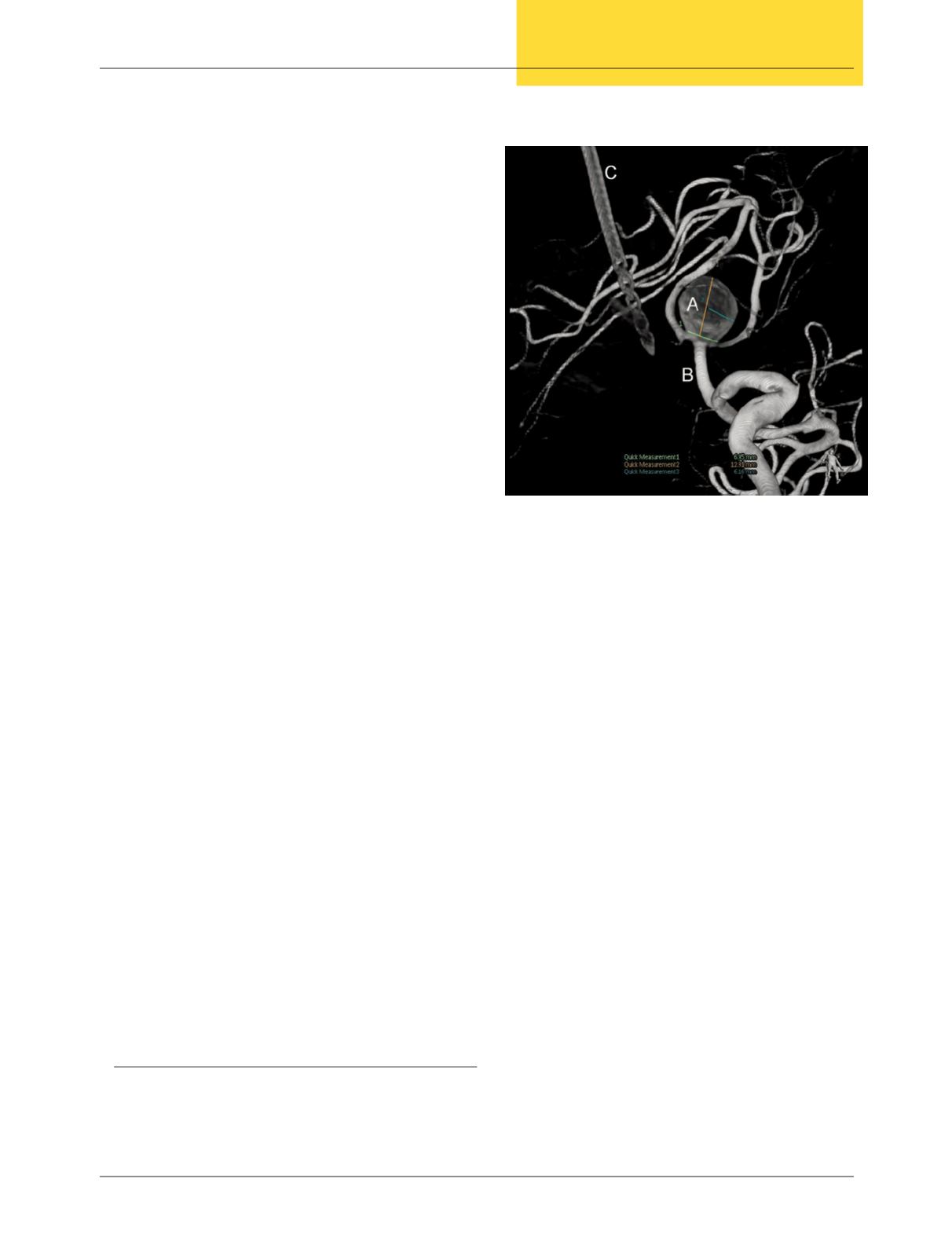
Figure 2: Detachable balloon
Detached balloon occluding the right ICA
(white hand)


